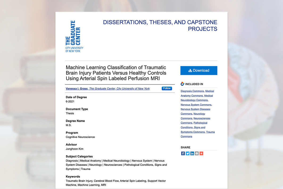
Graduate Neuroscience Research
RESEARCH, WRITING, & STRATEGY
The brain is arguably our most complex organ, acting as the command center for our thoughts, emotions, and behaviors. As part of my graduate research in Cognitive Neuroscience, I explored this complexity by investigating traumatic brain injury. My research involved gaining a deep understanding of brain structure and function, as well as neural activity, cognition, and behavior. I worked with top researchers, professors, and Ph.D. students at the City University of New York, collaborating on research strategy, analyses, and findings. Throughout this process, I made several key discoveries. One, in particular — that great research is really great storytelling — guided me as I wrote my master's thesis.
Background on Traumatic Brain Injury
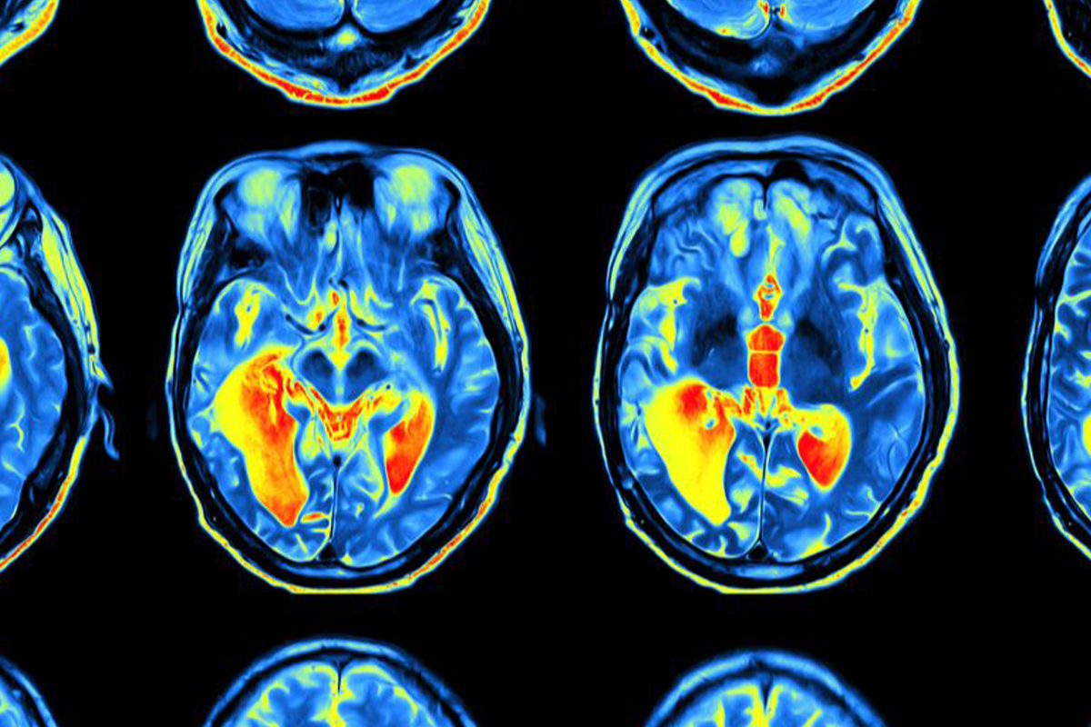
Traumatic brain injury is a major cause of death and disability worldwide. Common causes of traumatic brain injury include motor vehicle accidents, falls, and sports injuries. Unfortunately, traditional brain scans often don't give us the full picture of injury due to limitations in brain imaging technology. My research explored a novel approach to enhancing such brain scans with the overall goal of providing more accurate diagnoses and prognoses of recovery for patients affected by traumatic brain injury.
Literature Review
To lay the groundwork for my study, I undertook an in depth review of over 100 research papers across various domains of neuroscience. This rigorous exploration allowed me to comprehend the existing body of knowledge, identify trends, best practices and gaps in the current research, and develop a comprehensive understanding of the complexities surrounding traumatic brain injury.
Experimental Methods & Analysis
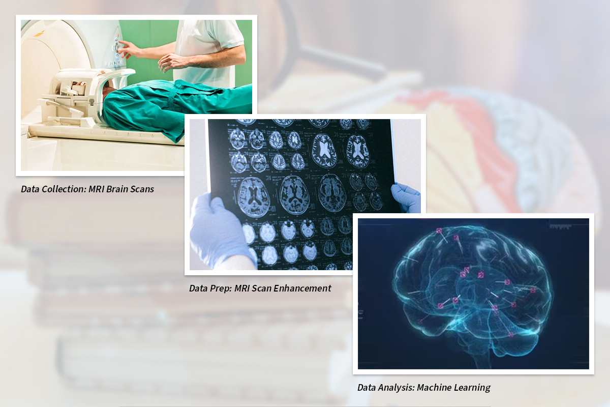
My research investigated brain scans from two groups: patients with traumatic brain injuries and healthy individuals who acted as control subjects. These scans were obtained using a specialized type of magnetic resonance imaging (MRI) technology. To enhance the quality of the brain scans, I applied brain imaging algorithms to make the scans cleaner and easier to analyze. For analyses, I utilized machine learning techniques to not only detect cases of traumatic brain injury but to also gain insights into the specific brain regions most affected by these injuries.
Results & Conclusions
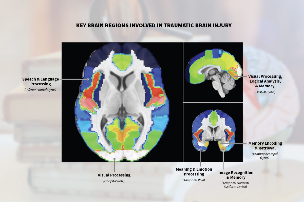
My research findings revealed that enhancing the quality of brain scans may lead to better detection of traumatic brain injury. Additionally, I identified potential correlations between traumatic brain injury and functional changes within key brain regions. Notably, these regions are known to play crucial roles in cognitive functions that are often impaired in individuals with traumatic brain injury.
Writing Process
My research writing process was highly iterative all while meticulously adhering to scholarly writing standards to ensure the credibility of my work. I dedicated significant attention to editing, proofreading, and fact-checking while referencing sources accurately and preparing my work for potential publication in scientific journals. Throughout the process, I regularly presented my findings and shared writing drafts with the lead researcher and fellow lab members. This collaborative approach deepened our collective understanding, ensured the reliability of the experimental methods and analysis, and developed a consensus on my results and conclusions.

Final Research Thesis
Overall, my research process was driven by a commitment to scientific rigor, collaboration, and continued learning. By implementing these strategies, I successfully produced a high-quality research thesis that made a meaningful contribution to the future study of traumatic brain injury.